Monitoring at MacMillan Wharf, Provincetown, MA

Monitoring at MacMillan Wharf, Provincetown, MA
Marine Invasive Species (MIS) Monitoring at MacMillan Wharf, Provincetown, MA.

Tuesday, December 31, 2013

Sea Stars at MacMillan Wharf

Grazing the Pilings and Harbor Floor

Sea stars are not usually seen on the floating docks, but this summer when I visited the marina at low tide, the docks had lowered on the pilings to the upper subtidal zone. On my last visit, I encountered several sea stars crawling over marine life on the pilings. From the morphology, coloration of the madreporite, and size, they appeared to be Asterias forbesi.
Sea stars prey on mussels and other species on the pilings and harbor floor. When feeding on a mussel, the sea star attaches its tube feet to each shell and exerts force to separate them slightly. A tug of war ensues, and only a small gap is sufficient for the sea star to insert a fold of its stomach and start digesting the body. When the mussel is sufficiently digested, the sea star brings its stomach back into its body with the food inside. The sea star has a well-developed sense of smell and can detect the odor of mussels and crawl towards them.
Sea stars are able to move around via a water vascular system of tube feet that act as suction cups to walk over the surface or pull prey into the mouth. The tube feet are located on the ventral surface and the water intake structure, called a madreporite, is located on the dorsal surface.

Docks and Pilings at MacMillan Wharf

Docks slide up and down on the pilings as the tide rises and lowers about 10 feet. Permanent growth of most algae and invertebrates can only be sustained below the low tide line.
Sea Star Near the Water Line of a MacMillan Wharf Piling
 
The center of the sea star is raised up as though it is feeding on a prey, or it is about to feed on the mussel wedged between two arms. When the sea star was lifted, it was not feeding on another meal, so perhaps is was headed towards the mussel.

Dorsal Surface of the Sea Star Asterias forbesi
Dorsal surface of the same Asterias showing the red madreporite and numerous spines covering the arms.

Ventral Surface of the Sea Star Star Asterias forbesi
Ventral surface of the same sea star showing the mouth and radiating rows of tube feet. The outline of the tube feet can clearly be seen in the shadow of the upper arm.
  
Sea Star Righting Itself after Being Place on its Ventral Surface
Sea stars can rapidly right themselves by flipping over using their tube feet and flexible bodies. This photo was taken 1-2 minutes after the photo above.
LINKS: 
Echinoblog:  Madreporite and Water Vascular System


Global Biodiversity Information Facility: GBIF.ORG: Asterias forbesi (Desor, 1848)

Saturday, November 30, 2013

Caprellid Amphipods Return to Docks

Caprella mutica back in Provincetown after Hurricane Irene

After Hurricane Irene passed through New England in late August, 2011, much of the growth on the docks at MacMillan Wharf was washed away, particularly delicate algae and invertebrates. One species that I failed to see in the following days and month was the amphipod Caprella mutica, also called the Japanese skeleton shrimp, which was observed fairly commonly on the docks earlier in 2011.  In 2012, Caprella had not re-established itself during my summer monitoring, but this year, groups of Caprella were found during July and August at several locations on the docks.   Most of the caprellids were attached to bushy hydroids and were not commonly observed on other filamentous (e.g., algae) or bushy species (e.g., bryozoa).   

My observations motivated a visit to the CZM MORIS website where species maps can be created for MIS species in previous years.  Confirming my previous observations, sighting of Caprella mutica decreased at different moriting sites from Narragansett Bay to Wells, Maine.   
 Groups of Caprella mutica living on branched hydroids
 
Populations of Caprella attached by their hind legs crawl over branched hydroids. Larger males and a few smaller females with mid-body brooding pouches can be identified. 
  
Caprella mutica MIMIC sightings in 2011 and 2012     
MORIS website-generated map of the locations where populations of Caprella mutica were found during invasive species monitoring in 2011 (top) and 2012 (bottom).  Caprella sightings decreased by about one-third.   No reports were made in 2012 in Narragansett Bay, Buzzards Bay, or Cape Cod Bay, and fewer sightings were reported in Salem Sound.  Website: MORIS: CZM’s Online Mapping Tool.  Credit: "Massachusetts Office of Coastal Zone Management (CZM), Executive Office of Energy and Environmental Affairs."
 
LINKS:
Harbor Watch:  Coastwatch 2012
"Caprella mutica (Caprellid amphipod a.k.a. skeleton shrimp) - Very common living on algae and bryozoa. These small amphipods look and act like miniature preying mantis.  They hold tight to surfaces and do not "swim around" like typical amphipods and shrimp." 
MORIS: CZM’s Online Mapping Tool
WILD Shores of Singapore: Workshop on Bryozoans and Hydroids: 29 April- 4 May, 2013.  "Tiny skeleton shrimps are commonly seen on some hydroids!"

Thursday, October 31, 2013

Floating Dock Mussel Beds

Provincetown Mussels and Ascidians

I've been monitoring floating docks at MacMillan Wharf in Provincetown for a few years now, and each season has been slightly different and yet broadly similar regarding the associations of different species.  One of the abundant species in the marina is the common blue mussel, Mytilus edulis.  It is abundant under the floats and variable in number on the sides depending on competition with other species and cleaning activities by marina personnel.  Each Spring, cleaned float surfaces provide opportunities for larval settlement. 

Mytilus normally lives on rocky shores in the intertidal zone attached to rocks and other hard substrates by strong, somewhat elastic structures called byssal threads.  These threads are secreted by byssal glands located in the foot of the mussel.  The mussels are firmly attached, but they have the ability to detach and reattach to the substrate allowing them to reposition themselves relative to their neighbors. They are usually found clumping together on wave-washed rocks, which helps hold the mussels firmly on the rocks against the force of the waves.

When the mussel larva first settles, it first secretes a thin shell and then develops an elongated foot with byssal glands. If the substrate is suitable, it will metamorphoses into a juvenile form and attach byssal threads. This attachment is a prerequisite for the foundation of the blue mussel population. On the sides of the Provincetown docks, large numbers of mussels will often settle on a clean surface and form masses of juvenile mussels which are striking because most of the individuals are about the same size, indicating that they settled around the same period of time.  These juvenile beds are apparently seasonal, because the beds do not mature over winter, and new beds of young mussels are seen the next Spring.  
 
Mussels can move slowly by extending a byssal thread, using it as an anchor and then shortening it.  A thread is formed by the foot by creating a vacuum at the contact site and secreting a foamy mixture of proteins into the formed chamber, producing sticky threads about the size of a human hair.
Mussels and ascidians frequent colonize together on floating docks, ropes, and fishing gear.  Ascidians compete for substrate, limiting colonization by mussels, and colonial ascidians will grow over the surface of the shell, limiting growth and food supply.  Co-colonization and competition of ascidians with mussels has had an impact on mussel aquaculture throughout the northeast.

Juvenile Mussels Beds on the Sides of Floating Docks
Mussel beds of young Mytilus edulis on the sides of floating docks at MacMillan Wharf, Provincetown, August, 2013.  Individual mussels averaged about 1-1.5 cm long.  In July, the mussels were covered by the colonial ascidian Diplosoma listerianum (which was extremely abundant), but most mussels were clean in August.  The soft ascidians were presumable removed by predation, an idea support by the observation in August of torn sections of Diplosoma colonies pulled off the substrate.  In these photos, small colonies of orange Botylloides violaceus and green-grey Diplosoma can be seen at the water surface, and two small colonies of Botryllus schlosseri (star tunicates) can seen deeper in the water in the top photo.  (Images in this post were taken with a 14 megapixel camera and can be enlarged without losing detail by zooming into the photo).
 
Mixed Floating Dock Communities of Mussels, Colonial Asicidans, and Algae
Botrylloides violaceus, green algae (Ulva), red algae (Neosiphonia harveyi), bryozoans (Bugula neritina), and other ascidians (Botryllus schlosseri, Diplosoma listerianum, Didemnum vexillum) form a colorful blend of species along with mussels.  Green-gray areas in top photo are Diplosoma.  Milky white areas in both photos are probably Didemnum.    
 
Mussels and Orange Colonial Ascidians Cohabitate Hanging Ropes 
Outside the juvenile beds, mussels grow and mature in small groups along with ascidians, especially Botrylloides violaceus.  Growth can become so extensive that the colony forms orange "hanging gardens."
 
LINKS:
A Snails Oddessy:  Learn about Mussels. Anchoring.
Van Winkle, W.  Effect of environmental factors on byssal thread formation.  Marine Biology 7: 143-148, 1970. 
Lane, DJW, AR Beaumont, and JR Hunter.  Byssus drifting and the drifting threads of the young post-larval mussel Mytilus edulis.  Marine Biology 84: 301-308, 1985.

Monday, September 30, 2013

Striped Anemone's Life in Wellfleet

Surviving Adverse Tidal Conditions
 
After I first started monitoring Wellfleet Marina in 2011, I wrote a post on the striped anemone, Diadumene lineata, on docks in the North Harbor, noting that few other species were found (see December 2011 post).  A monitoring study on MIS species along the New England coast reported that Diadumene was extremely tolerant to extremes in temperature, salinity, and water quality (Pappal et al, 2003).  Interestingly, temperature and salinity readings in Wellfleet during the summer of 2011 appeared normal, similar to those in Provincetown.  I did notice, however, that the turbidity of the water was much higher than that in Provincetown, and visibility was greatly reduced.

The following year, I learned more about the harbor when I coincidentally visited the harbor about 30 minutes before low tide.  The North Marina was already drained of water, and, in the South Marina, the receding water under the docks was rapidly disappearing.  I realized that the twice-daily exposure at low tide and high sediment levels were probably the most significant contributing factors to the distribution of species (see additional images in Footer at bottom of Blog).

High tidal variation is characteristic of the Gulf of Maine, and in the North Harbor, the shallow bay is filled and emptied during each tidal cycle. This variation has a positive effect on marine life (e.g., oyster beds) in that it brings in fresh seawater twice a day.  At the highest tides, the harbor is filled and Duck Creek is a shallow bay.  At the lowest tide, Duck Creek is drained and the marina is transformed into a mudflat.  Tidal variation in the summer averages over 10 feet, and this July, the tidal variation peaked at over 14 feet.  
 
On the north marina, Diadumene lineata, a yellow sponge (keyed as Halichondria bowerbankia), and an occasional oyster have most of the floating docks to themselves.  At high tide, sediment in the water produces a layer of sediment and organic matter on the sides of the floats.  At low tide, the floats sink into the mudflat.  This adds an additional layer of dark mud to the float.  Most of the anemones are in the upper layer near the waterline.     
 
On the South Marina, most of the docks are seasonal, and Molgula, algae, and distinctive color variations of Botryllus schlosseri form the dominant species on the sides of docks.  Other species that are commonly observed are sea lettuce Ulva, filamentous green algae, an occasional Codium, and branched red algae such as Neosiphonia harveyi

Satellite Views of Wellfleet Harbor at High and Low Tide
Tidal variation in Wellfleet Harbor taken from different satellite images of Wellfleet.  Left, high tides.  Right, mid-to-low tide.  At the lowest low tides, the entire north estuary is drained. 

View of the North Wellfleet Harbor at Low Tide
Evening low tide in the Duck Creek estuary on the North Side of Wellfeet Marina, September 16, 2012.  The entire area is drained and converted into a mud flat of dark brown, wet sediment.  At low tide, the docks rest partially submerged in sediment. This year, the marina was monitored at high tide and will be henceforth.
 
Diadumene Habitat on the Side of the Main Dock in the North Marina 
Digital photos and enlarged details of Diadumene lineata on the North Marina docks (August, 2013).  Diadumene lives in social groups distributed along the length of the docks right below the water line.  Wellfleet was monitored at high time this summer.  The floats were stratified into three regions: the upper, dry float above the water water (bottom of top image), a light brown zone containing sediment below the water line that is exposed at low tide, and a dark brown zone zone that sinks into the dark sediment at low tide (top of top image).  Most of the anemones live in the light brown region, but a few individuals can also be found in the dark brown zone.    
 
LINKS:
Pappal, A, J Pederson, JP Smith.  Marine Invaders in the Northeast, 2003.
In this study, Diadumene was commonly found at Marinas in Naragansett Bay and Buzzards Bay and at certain locations in the Gulf of Maine. 
Harbor Watch:  Striped Anemone at Wellfleet Harbor.  Features stereomicroscopic views of collected anemones.     

Saturday, August 31, 2013

Codium: Biology of a Marine Invasive

Multinucleated Single Cell Green Alga


Codium fragile is a large, dark green macroalga with one to several, thick upright branches arising from a broad, spongy basal disc attached to the substrata.  The cylindrical branches are dichotomously branched and arise from a juvenile phase having both prostrate and erect branches.  Fronds are generally annual, dying back in the Winter and arising from the perennial basal portion in the Spring.
 
The branches are constructed of interwoven coenocytic filaments, all derived from the same germ cell. Like Bryopsis (January 2013 post), there are no cell walls to separate nuclei or individual cells.  However, the structural organization of Codium is distinctly different from Bryopsis and other coenocytic green algae.  Each multinucleated branch is composed of a network of fibers that orient pointed cellular extensions, called utricles, toward the outside.  The utricles of Codium fragile have a thorn-like projection that is absent in other species. The utricles are packed tightly, side by side, creating an outer layer surrounding the filaments. 

Codium has been grown in the laboratory to study differentiation and regeneration.  In early studies, cultures were primarily composed of dissociated branched, coenocytic filaments. The typical growth form of upright branches with utricles did not develop.  With further research, techniques to grow mature filaments and branches were developed.  Branched algae were developed from heterotrichous juveniles when cultures were agitated on a shaker. The shear forces created by mechanical agitation were essential for both initiation and maintenance of upright branches.  Using aquaculture techniques, Codium has been grown in Korea from regenerating isolated utricles and medullary filaments in a step-by-step manner (see LINK below).   

Codium fragile
 Codium fragile growing on the side of a floating dock at MacMillan Wharf, Provincetown, MA. Photo was taken during a monitoring session in August, 2013. Codium typically grows along the water line in the same location as Ulva, Enteromorpha, and other green algae.

The Life Cycle of Codium
Diagram of the life cycle of Codium showing upright thallus, cross section through a branch, utricles, gametes, and dichotrichous germling.  The cross section through the branch (fertile thallus, bottom left) shows the central filamentous multinucleate cell and the outer utricles.  Male and females gametes fuse together to form the zygote, which then develops into a germling that grows to form the holdfast and diploid thallus. 

Codium fragile Internal Structure
 
Diagram of a cut Codium branch showing central filaments and the outer utricles.

Microscopic Structure Codium fragile utricles
 Left, diagrams of Codium utricles showing fusiform gametangia forming at the side of the utricle. Microscopic image of Codium utricles.
 
LINKS:
Dixon, HH.  Structure of Codium.  Ann Bot 11: 588-590, 1897.   
Ramus, J.  Differentiation of the green alga Codium fragile Amer J Bot 59: 478-482, 1972.
MH Yang, G Blunden, FL Huang.  Growth of a dissociated, filamentous stage of Codium species in laboratory culture.  J Applied Phycol 9:1-3, 1997.
Hwang, EK, JM Baek, and CS Park. Cultivation of the green alga, Codium fragile (Suringar) Hariot, by artificial seed production in Korea. J Appl Phycol 20: 469-475, 2008.
Aquaculture of Codium fragile.  b-d) filaments are grown on coiled fibers for about 6 weeks.   e) with continued culture, erect thalli develop. f-h) once erect thalli develop, the fibers are coiled around a larger culture rope.  Small branched plants are formed by 5 months (g) and mature growth is achieved by 7 months (h). 
Encyclopedia of Life (EOL):  Codium fragile
Natural History of Sado Island, Japan:  Codium fragile.
Flora of South Australia:  Codium fragile (Suringar) Hariot
Natura In Neustria:  Codium fragile (algue verte)
Taxonomic Toolkit For Marine Life of Port Phillip Bay http://portphillipmarinelife.net.au
Codium on the beach at Port Phillip
Montery Bay Aquarium Research Institute: Codium setchellii. Structure of C. setchellii compared to C. fragile.

Wednesday, July 31, 2013

Summer Monitoring in Provincetown

Time to Enjoy the Scenery

View of the study area - two public docks off the East side of MacMillan Wharf.  At the end of the afternoon, dark clouds came in from the Northeast creating a picturesque effect.

The North side of the first dock is used for small boats and has a few wood-framed floating docks maintained by local organizations.  The South side of this dock features berths for commercial fishing vessels.  

 A view from the Wharf looking Southeast towards the second dock.  Both sides of the dock have berths for fishing vessels.

Sunday, June 30, 2013

Coast Watch - 2013

Expectations for the Season

It's another monitoring season and plans are underway for assessing marine invasive species in Provincetown and Wellfleet this year. Looking back at the findings from the last two years raises somes questions about this year's marine growth.  Each Spring, the sides of docks usually have areas of clean substrate that are available for settlement.  Both invasive and native species compete for these sites.  

Ascidians are one of the most competitive groups of species that attach to the docks.  In the Gulf of Maine, the colonial species Diplosoma, Didemnum, Botryllus, and Botrylloides, and the solitary ascidians Styela, Ascidiella, Ciona, and Molgula complete with other groups such as mussels, bryozoa, and algae. Last year, Diplosoma was a major colonizing ascidian of clean substrates, co-colonizing with Botryllus and Botrylloides on the sides of docks below the water line.  Ascidiella has not yet established a foothold in Provincetown, although it is common in other parts of the Gulf of Maine (www.salemsound.org).  In contrast, Molgula which occupies a similar ecological niche to Ascidiella, is common and may have a competitive edge in settlement due to the large number of individuals producing larvae.

It will be interesting to see whether two MIS crustaceans make an appearance and, if so, how abundant they will be.  Caprella mutica was not recorded in Provincetown last year although it was abundant in 2010 and early 2011, and a few Palaemon elegans were seen for the first time in Provincetown during the summer of 2012 living among schools of Palaemonetes pugio.

I find that a visit to the docks is always enhanced by searching for hanging ropes, submerged boating gear, buoys, or improvised objects such as automobile tires that may be attached to the sides of docks.  Ropes hanging from the docks usually show variation in algae and invertebrate species distribution with depth due to light and temperature factors.  Juvenile green and Asian crabs, usually around the size of a dime or nickel, are typically seen during monitoring sessions crawling over species on the docks, on ropes, or living in the protection of attached structures on the dock. The photos below show a few examples of previous years findings.
 
Public Docks at MacMillan Wharf 
The main docks rest on large concrete-covered styrofoam floats whereas most of the side docks rest on modular commercial floats composed of expanded polystyrene cores enclosed by a black polyethylene shell. 
 
Codium Green Algae and Colonial Asicidians on a Hanging Rope
Part of a light weight rope that looped from one dock to another.  Near the water line, Codium covered a short strand of rope tied to a dock.  Orange Botrylloides violaceus covered another section of the rope that was hanging deeper in the water.  A few small specimens of Ulva sea lettuce can also be seen along the rope.  

Small Mytilus edulis Mussels on a Nautical Rope 
Mussels are another species that colonizes docks and ropes near the waterline.  When larvae settle at the same time, a cohort of uniformly sized mussels is formed.
 
Heavy Colonization of a Hanging Rope by Colonial and Solitary Ascidians
Didemnum, Botryllus, Botrylloides, Molgula, and other invertebrates grow over each other and entangle eel grass and other debris to form large masses on a hanging rope.
 
Spider Crabs Visit the Main Dock of MacMillan Wharf
Two young Libinia emarginata spider crabs were found hanging out together in a protected area under the dock.  One individual had a small colony of Botrylloides violaceus growing on its back.  This was a rare treat because green and Asian crabs are usually the only crab species that are seen on the docks.

Friday, May 31, 2013

Club Tunicate First Described by William A Herdman in 1882

Collected on the Voyage of the HMS Challenger
 
The Challenger expedition of 1872–76 was a scientific exercise that made many discoveries to lay the foundation of oceanography. The expedition was named after the mother vessel, HMS Challenger.  Prompted by Charles W. Thomson of the University of Edinburgh, the Royal Society of London obtained the use of Challenger from the British Royal Navy and in 1872 modified the ship for scientific work, equipping it with laboratories, workrooms, and storage space.
 
To enable the ship to probe the ocean's depths, the Challenger's guns were removed and its spars reduced to make more space available. Laboratories, extra cabins and a special dredging platform were installed. It was loaded with specimen jars, filled with alcohol for preservation of samples, microscopes and chemical apparatus, trawls and dredges, thermometers and water sampling bottles, sounding leads and devices to collect sediment from the sea bed and great lengths of rope with which to suspend the equipment into the ocean depths. Because of the novelty of the expedition, some of the equipment was invented or specially modified for the occasion. Under the scientific supervision of Thomson, the Challenger  sailed from Portsmouth, England, on 21 December 1872, and travelled nearly 70,000 nautical miles surveying and exploring the oceans.
 
The final results of the research were published as the "Report of the Scientific Results of the Exploring Voyage of H.M.S. Challenger during the years 1873-76". Among many other discoveries, the report catalogued over 4,000 previously unknown marine species. One of these species was the as-of-yet un-named Styela clava. William A. Herdman described and illustrated the Tunicates from the expedition publishing several reports in 1882 on simple, compound, and pelagic Tunicates.  
 
HMS Challenger
British Natural History Collections image of the Challenger, 1872
 
Route of the HMS Challenger
Styela clava was collected off the coast of Japan during explorations in 1874.
 
Herdman noted that S. clava appeared to be a rather common species of Styela in Japanese seas. At the time, there were about twenty specimens of it in the British Museum collection, which were brought to England from Japan, and there were also some specimens from the same locality in the Liverpool Free Public Museum. The species, however, appeared to have been never described or illustrated. The text described the characteristic features of the species and illustrations showed the external appearance and structure of the brancial sac.
 
Herdman wrote that the species was club-shaped with an pyriform body supported on a stalk of variable length that stood erect and was not compressed. The branchial sac of the specimens had four narrow folds upon each side. The internal longitudinal bars were numerous, about nine on a fold and twelve in the interspaces.  The meshes were transversely elongated and each contained six stigmata.

Original Illustration of Styela clava
Figure 9, external appearance of 2 specimens of Syela clava, WA Herdman, 1882
 
Branchial Sac and Stigmata of Styela clava and several other species of Styela
Plate XIX from "Tunicata: The Report of the Voyage of the HMS Challenger".
Styela clava, Fig. 9 and 10. Figure 10, part of the branchial sac from the inside showing branchial bars and stigmata.

LINKS:
Wikipedia Website:  Challenger expedition
National Oceanic and Atmospheric Administration (NOAA): Ocean Explorer.  Then and Now: The HMS Challenger Expedition and the “Mountains of the Sea” Expedition.
University of Kansas Natural History Museum, Division of Invertebrate Zoology. Challenger Expedition (1872-1876).
Isle-of-Man Webpage:  William Abbott Herdman, 1858-1924
Herdman, WA. Report of the Scientific Results of the Voyage of H.M.S. Challenger During the Years 1873-76. Zoology Part XVII. Ascidiæ Simplæ. Proceedings of the Royal Society of Edinburgh, 1882.
19thcenturyscience.org:  Comprehensive Overview of the Findings of the Challenger
19thcenturyscience.org:  Tunicata: Ascidiae Simplae.
19thcenturyscience.org: Tunicata: Ascidiae Compositae.

Title Page
 
Styela clava, Pages 158-159 

Tuesday, April 30, 2013

Japanese Tsunami Floating Docks

Asian Species Arrive on West Coast Shores

Recently, it has become a regular occurrence that another piece of debris with attached marine species arrives on the shores of Washington and Oregon as a result of the 2011 Japanese tsunami.   These reports are fascinating from several perspectives: dispersal of marine species following natural events, a glimpse at Pacific species, and the effect of transatlantic travel on species distribution.  Four large docks were washed away from Misawa, Aomori Prefecture, Honshu.  One was found in Japan, two have arrived in the United states, and one is still unaccounted for.  It will be interesting to learn how this process unfolds in comparison to the much different process of marine species on ships that travel from port to port. Their time at the shore was limited because they took a few days to permanently wash ashore and because they were immediately cleaned and removed from the area.  These docks had their greatest potential to impact the open coast because marinas and bays are relatively few in number or protected from the open coast.
  
The first dock arrived in Oregon on June 4th, 2012, at Agate Beach, Newport. This was a large dock densely covered with attached algae and marine invertebrates.  Coincidentally, Newport is the home of the Hatfield Marine Science Center of Oregon State University and The Oregon Coast Aquarium.  It was a ideal situation for documenting the species and studying their impact on the surrounding areas. Since then, another floating dock was reported washing ashore December 16-18, 2012, in a remote location along the Washington coast near Mosquito Creek on the Olympic Peninsula.  The docks had followed the predicted path of debris from Japan based on prevailing currents, winds, and storms, but they arrived at North America slightly ahead of schedule.  
 
Several research groups and experts are participating in identifying the species on the docks, especially the Agate Beach dock which has over 150 species and was densely covered.  Each list is posted on the web and is periodically updated (see LINKS).  It will be worth following during the upcoming months to see what species appear as the story unfolds.  Several Gulf of Maine MIS species were also seen on the docks including Styela, Didemnum, Grateloupe, and Caprella.  Some invasive species were also seen that are already on the West Coast, including the bryozoan Watersipora subtorquata and the Mediterranean mussel Mytilus galloprovincialis.  Several encrusting and upright bryozoans were identified.  Cryptosula pallasiana and Watersipora subtorquata are encrusting forms with distinctly different enclosures from Membranipora or Electra (see links and images below), and Tricellaria sp and Scruparia sp are beige branching forms structurally different from our Bugula species.  
 
Perhaps one of the most interesting findings from my perspective was the presence of numerous stalked, pelagic gooseneck barnacles that settled on the dock during the trip across the Pacific. Pelagic barnacles are normally found attached by their flexible stalks to floating timber, the hulls of ships, piers, pilings, and seaweed. Lepas anatifera has a cosmopolitan distribution and is found in tropical and subtropical seas worldwide. Because it is attached to floating objects carried by oceanic currents, it is ofter found in colder seas where the waters are too cold for them to reproduce. It will be interesting to learn what other species were acquired during the trip, whether species that were established on the docks were lost or diminished in density during the journey, or whether other species became more dominant due to more advantageous conditions.
 
Location of the Floating Dock on Agate Beach, Newport, Oregon
Satellite view of Western Oregon over Newport, Oregon showing location of the Misawa dock represented by the yellow rectangle.  Red star:  location of Hatfield Marine Science Center and Oregon Coast Aquarium.  

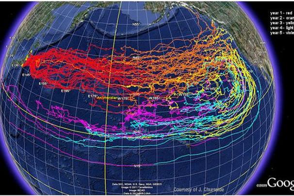
Predicted Path of Debris From the 2011 Tsunami Around the North Pacific
NOAA's OSCURS (Ocean Surface Current Simulator) is a numeric model for ocean surface currents that predicted the movement of marine debris generated by the Japan tsunami.  Debris drifted more rapidly than anticipated, reaching Alaska first and then Oregon in 2012.  The model shows that once the debris reaches the eastern Pacific, it can either pass through the California current and reach the coast, or get caught in the current and travel south.  More southerly, the current turns west and debris moves west traveling back to the west Pacific.  Time sequence progresses as follows:  Red, Orange, Yellow, Turquoise, Magenta.

Japanese Floating Dock on Agate Beach, Newport, Oregon
The Misawa dock came to its resting place on a long sandy beach.
(Credit: David Appell)

Marine Growth on the Dock at Agate Beach
 An initial survey found a variety of species of barnacles, starfish, urchins, anemones, amphipods, worms, mussels, limpets, snails, solitary tunicates and algae. This section shows numerous mussels  (Mytilus galloprovincialis ) and brown algae. 
M. galloprovincialis also occurs on the West Coast of America. 

Chart of Marine Organisms on the Agate Beach Oregon Floating Dock
Chart of several of the species found on the Agate Beach Floating Dock.  A large diversity of species was found. The solitary tunicate on the left looks like a pair of Styela clava.  Encrusting and pelagic barnacles are shown in the upper right.
 
Japanese Floating Dock near Mosquito Creek, Olympic Peninsula, WA

Pelagic barnacles settled on the dock during transport across the Pacific.  The side looks nearly covered with the single species, Lepas anatifera

Lepas anatifera Pelagic Barnacles
 
Body divided into two parts: the capitulum which bears the body of the animal and a flexible stalk called the peduncle.  Capitulum has five large calcareous plates.  Plates on the capitulum are smooth or at most finely marked. Upper left, specimens with the cirri withdrawn showing external features - the plate-covered capitulum and the leathery,brown peduncle Upper right, collected specimens with cirri showing morphological diversity.  Bottom, underwater image showing orange tissue-lined pearly white plates and fully extended cirri ready for feeding.

LINKS:
Oregon
Oregon State Japanese Tsunami-Generated Floating Docks Website.  Gives updated information on the research and species list.  http://blogs.oregonstate.edu/floatingdock/
ANS (Aquatic Nuisance Species) Task Force.  http://anstaskforce.gov/Tsunami.html
Oregon State Tsunami Debris Hotline: Huffington Post ongoing dialogue of reports and replies.
Agate Beach Tsunami Dock Species List.  Gives updated species information.
Tsunami Floating Dock
Preliminary Species List : As of January 1, 2013 
Marine Organisms Found Living on a Floating Dock from
Misawa, Aomori Prefecture, Japan Dislodged by the 2011 Tōhoku Earthquake and Tsunami and Washing Ashore on June 4, 2012 at Agate Beach, Lincoln County, Oregon
------------

Washington
Washington Tsunami Debris: Olympic Peninsula Dock.  Huffington Post Report.
Washington Coast Tsunami Dock.  NOAA web report.
Species List Washington Misawa Tsunami Floating Dock.  Gives updated information. 
Tsunami Floating Dock: Misawa 3
Olympic National Park
(JTMD-BF-8)
Species List: As of January 15, 2013
Marine Organisms Found Living on a Floating Dock from
Misawa, Aomori Prefecture, Japan, Washed Out to Sea on March 11, 2011,and
Washing Ashore December 16-18, 2012 near Mosquito Creek, Jefferson County,
Olympic National Park, Washington
------------
Bryozoa
Encyclopedia of Life (EOL): Cryptosula pallasiana. Overview of the species.
The Exotics Guide:  Non-native Marine Species of the North American Pacific Coast.  Watersipora subtorquata.  Detailed description of the taxonomy, structure, and distribution of the species.
Watersipora subtorquata is an invasive species that has also spread to the US Pacific coast. Top, View of an orange colony from San Francisco Bay (Luis A. Solórzano).  Bottom left,   Closeup of Watersipora subtorquata zooecia showing the black opercula and the fine black lines where the zooecia join (California Academy of Sciences).  Bottom right, SEM image showing fine structural detail.
Encyclopedia of Life (EOL): Tricellaria. Genus overview. 
Tricellaria inopinata D'Hondt & Occhipinti Ambrogi, 1985
Encyclopedia of Life (EOL): Scruparia. Genus overview.
Scruparia chelata Linnaeus, 1758, is a white colony with creeping stolons
and upright chains of zooids
------------
Mussels:
Wonham, NJ. Mini-review distribution of the Mediterranean mussel Mytilus galloprovincialis (Bivalvia: Mytilidae) and hybrids in the Northeast Pacific.  J Shellfish Res, 23: 535–543, 2004.
------------
PelagicBarnacles:
MarLINCommon Goose Barnacle:  Lepas anatifera
EOL Encyclopedia of Life:  Lepas anatifera.
WoRMS World Register of Marine Species. Lepas anatifera Linnaeus, 1758.
------------
Recent News:
Japanese Fish Survive 5,000-Mile Trip across Pacific in Tsunami Boat.  Blue and white striped beakfish survived in the hull of a boat.