Monitoring at MacMillan Wharf, Provincetown, MA

Monitoring at MacMillan Wharf, Provincetown, MA
Marine Invasive Species (MIS) Monitoring at MacMillan Wharf, Provincetown, MA.

Saturday, April 28, 2012

Botryllus: Burgundy Color Variants

At the beginning of the season, the typical star-shaped Botryllus schlosseri color variants with a burgundy body and white inter-siphon pigment bands forming an actual white star was one of the most prevalent color variants.  But as the summer progressed, the number and variations of color variants increased, including the burgundy colonies.  Their appearance in daylight or under incandescent light varied depending on substrate (light green sea lettuce, dark green Codium, brown algae, red algae, mussel shell, Styela) and background setting (on the dock, in collection containers, or on a black or white microscope stage). In this post, I show a few of the burgundy color variants collected from Provincetown and Salem Sound during the summer.

   Burgundy Botryllus schlosseri Variant on Green Algae - White Star
Botryllus schlosseri on Ulva, MacMillan Wharf, Provincetown, MA, September, 2011.  Green back-lighting through the algae clearly outlines the zooids.   White pigment cells form a pair of curved bands between the branchial and excurrent siphons giving the bands a daisy-star appearance.  Stereozoom 1.0 x 10x.  

Burgundy Variants with Inter-siphon White Shield

 
Botryllus schlosseri on Codium, MacMillan Wharf, September, 2011. White pigment cells form a shield-shaped pattern filling the space between the branchial and excurrent siphons of the cluster. The zooids also have a tiny patch of white cells on the opposite, outer side of the branchial siphon at the anterior end of the endostyle.  Othewise, the branchial siphons are burgundy like the rest of the zooid body. Stereozoom 3.0 x 10x.

 Burgundy Variant on Red Algae, White Branchial Crescent with Atrial Ring
 
Nine-zooid cluster of Botryllus schlosseri on red algae, MacMillan Wharf, October, 2011. Scattered white pigment cells encircle the orange-colored branchial siphon, more heavily on the outer side of the cluster. The common excurrent siphon is outlined by a dashed ring of white pigment cells formed by a short, white stripe at the atrial opening of each zooid.  Stereozoom 1.5 x 10x.

Ampullae of Burgundy Variant on Red Algae - White-Capped
 
Same colony of Botryllus schlosseri on red algae as shown above.   The zooids share a common vascular system that consists of blood vessels and enlarged club-shaped vascular ampullae along the periphery. The ampullae have the capacity to reconstruct the colony by vascular budding if all the zooids are lost.  Top, ampullae define the edge of the colony and separate clusters of zooids.    Bottom, white pigmented cells concentrate at the distal ends of burgundy ampullae like snow-capped boulders. 

Burgundy Variant on Red Algae - Diffuse White Branchial Stripe

Botryllus schlosseri on red algae, MacMillan Wharf, September, 2011.  White pigment cells form a short stripe through the branchial siphon above the endostyle and dorsal lamina.  Pigment cells also lightly speckle the body of the zooid.  Branchial siphons are orange-colored. Top, two 7-zooid and 10-zooid clusters.  Stereozoom 2.0 x 10x.  Bottom, close-up view of branchial siphons and diffuse white stripe.  Stereozoom 3.0 x 10x.  

LINK:

Friday, March 30, 2012

Identifying the Native Encrusting Bryozoan

Electra pilosa

Electra pilosa is a native encrusting Bryozoans found in the Gulf of Maine co-habitating with the cosmopolitan, MIS species Membranipora membranacea. Both of these encrusting colonies have a lacy appearance and are typically found on brown algae such as Laminaria sharing space on the same blade. Upon inspection, established colonies may look similar but they can be distinguished from each other by specific differential features. 

Colony growth pattern is an important clue to differentiating Electra from MembraniporaElectra in its first stages of growth forms star-shaped colonies whereas Membranipora grows in a uniform, radial pattern (what Silén called "unitary multiserial", see LINK below).  In contrast, the colony of Electra is separated into sectors divided by radial axes that extend beyond the margins of the colony forming a multi-pointed star (what Silén called "composite multiserial" growth).  Each sector has a central growth axis composed of 2 or more parallel rows of rectangular zooids (approximately 0.33 by 0.5 mm in dimensions) flanked on each side by wings of oval or ovate zooids which fill in the area between the axes. The side walls of the calcified enclosure do not directly abut each other like in Membranipora (see February 12, 2012 post), but are separated by a translucent, calcareous surface membrane. Each enclosure is surrounded by several spines that vary in length. However, the median, proximal spine is larger than the rest and can be easily seen with a magnifing lens. 

The bottom line when identifying encrusting Bryozoans in New England is to check for growth pattern (composite vs. uniform), boundary morphology (jagged vs. smooth), zooid skeletal structure (ovate vs. rectangular), arrangement of spines (larger pointed, proximal spine vs. 6 short, blunt spines, 4 at each corner of the rectangular enclosure) for Electra vs Membranipora, respectively.  

Growth Pattern of Electra vs. Membranipora
Comparison of growth pattern of Electra vs. Membranipora in small colonies where the axial structure of Electra is most pronounced.  Modified image from Silén, 1987 (see LINK below).  

Axial Growth Pattern of Electra pilosa
Outer border of Electra pilosa colony growing on Laminaria collected from Hawthorne Cove, Salem Sound, MA.  The colony illustrates composite multiserial growth.  At the bottom, two axial strands of rectangular zooids curve toward each other encircling inter-axial ovate zooids. At the top, several axial strands join each other to form a single 5-zooid wide strand.   Stereozoom 1.0 x 10x objective. 

Calcareous Enclosures of Electra pilosa
Electra pilosa zooids showing axial (left and right) and inter-axial zooids (center) with conspicuous proximal spines and translucent calcareous membranes between walls of the enclosures.  Stereozoom 3.0 x 10x objective

LINKS:


Sunday, February 12, 2012

MIS Encrusting Bryozoan

Membranipora membranacea
Bryozoans are tiny colonial organisms characterized by the presence of a tentacled feeding structure called a lophophore that filters food particles out of the sea water and transfers them to the mouth. The tentacles of Bryozoans differ from those of anemones and corals in that they contain a body cavity and the anus is located outside the circle of lophophore tentacles.  Encrusting Bryozoans in the Gulf of Maine include the cosmopolitan Membranipora membranacea and the native Electra pilosa.  Encrusting colonies have a lacy appearance and are typically found on brown algae such as Laminaria species but are also found on red algae and solid flat surfaces in marinas.

Membranipora forms mat-like calcareous colonies of small, closely packed zooids living inside a rectangular sheath that secretes a protective, calcified enclosure.  Each rectangular compartment is composed of lateral calcified walls and ventral uncalcified bands, which provide flexibility.   A membrane completely roofs the space between the supporting side walls. The enclosure has an orifice through which the lophophore extends into the water column for feeding. In Membranipora, the orifice is covered by an operculum

Membranipora membranacea Growing on a Blade of Laminaria
A small colony of Membranipora membranacea growing on a blade of the brown algae Laminaria longicruris collected from Beverly Marina  The point of origin (the ancestrula) is in the upper left quadrant.  Membranipora is characterize by rectangular enclosures and a smooth edge lacking zooids along the growing colony edge.  1x zoom x 10x objective.

Smooth Growth Zone at the Edge of Membranipora membranacea Colony
Growing edge of Membranipora membranacea lacks zooids and provides a smooth edge for excurrent water flow away from the colony.  1.5x zoom x 10x objective.

Diagram of an Encrusting Bryozoan: Retracted (left) and Feeding (right)
Diagram of two encrusting zooids, one retracted and one with the lophophore extended.  The lophophore circulates water through the tentacles, filters food particles, and transfers them by ciliary motion to the mouth.  Because the zooids are closely packed, the lophophores of the colony work cooperatively to circulate water through tentacles and to remove waste and unwanted particles.  When disturbed, the retractor muscles pull the zooid and lophophore into the safety of the enclosure. From www.earthlife.net - bryozoa.

Membranipora membranacea Rectangular Zooid Chambers
Calcified rectangular walls of the colony of Membranipora membranacea.  The units are arrange in staggered rows that give the structure additional strength.  Short tubercles (spines) occur at the corners of the rectangular enclosures, 6 spines per enclosure.  4x zoom x 10x objective.

Links:
Beyond the Human Eye
WoRMS: Membranipora membranacea

Web Photo, showing Colony Enclosures by Scanning Electron Microscopy:

Membranipora organization by SEM Dennis Gordon

Web Photo Favorites, showing colony formation:
Marney Pratt's Home Page, Duke Univ

Colony in Culture showing lophophores Ann-Margret Amui-Vedel

References on Feeding and Water Flow:
1. Pratt, MC. Effect of zooid spacing on bryozoan feeding success: is competition or facilitation more important? Biol. Bull. 207:17-27, 1004.
2. Pratt, MC. Consequences of coloniality: influence of colony form and size on feeding success in the bryozoan Membranipora membrancea. Mar. Ecol. Prog. Ser. 303: 153-165, 2005.
3. Von Dassow, M. Effects of ambient flow and Injury on the morphology of a fluid transport system in a Bryozoan. Biol. Bull 208:47-59, 2005.
3. Winston, JE. Current related morphology and behavior in some Pacific coast Bryozoans. In Advances in Bryozoology: Proceedings of the 4th International Conference on Bryozoa, GP Larwood & MB Abbott, eds. Systematics Association Special Volume, 13:, 247-268, 1979.

Saturday, January 21, 2012

Diplosoma Ascidian Up Close

Diplosoma listerianum is a cryptogenic species of almost cosmopolitan distribution that is found from the low-tide mark until about 80 m deep and is a close relative of Didemnum vexillum.  Both species have rapidly spread throughout New England waters and have become dominant species in many localities.  They are members of the same ascidian family, the Didemnidae, and and have a similar zooid body structure but differ primarily by the presence of stellate spicules and a coiled sperm duct in Didemnum.  The lack of spicules gives Diplosoma a smooth, gelatinous feel to it, whereas Didemnum has a tougher, slightly calcareous surface.  By eye, the two species would rarely be confused because they differ dramatically in color.  Dilosoma is an inconspicuous green-grey whereas Didemnum is a bright tan or cream color.  The feel of Diplosoma comes into play when trying to determine the identity of an amorphous green gelatinous mass, particularly when feeling the out-of-view but within-reach under-surfaces of docks. 

Diplosoma listerianum Colony showing Distinctive Green-Grey Color.
Numerous, small openings in the surface of the colony are incurrent branchial siphons of individual zooids.  Less frequent, large openings are common, excurrent apertures.  
Diving photograph by Rokus Groeneveld, Diverosa.com

During the monitoring season, Diplosoma was seen throughout the summer in Salem Sound but did not make its first appearance at the Provincetown Docks until August and continued to spread through September. I never saw it on the sides of docks in Wellfleet.  Earlier in the season, most of the Diplosoma specimens I brought to the lab were not very photogenic because of their dark color, lack of contrast, or poor viability in standing sea water.  However, in September, I collected a colony growing on red algae that I kept in replenished sea water until it was examined in the lab the next morning.  This colony showed beautiful light-blue structural features and white pigmented cells in the surface that glowed like stars in the sky.  The zooids, which are only about 2 mm long, afforded great views of the branchial siphons and stigmata in the branchial sac.   

Diplosoma listerianum on Red Alga Glows Light Blue by Stereomicroscopy
This view is from above, looking almost directly down on the branchial siphons, which have 6 lobes and up to 24 thin branchial tentacles of variable size.  The branchial sac bears 4 rows of stigmata, which can be clearly seen through the opened branchial siphon arranged in a ring around the opening. 

Diplosoma  listerianum Zooids showing Rows of Light Blue Stigmata
 This view is looking at a slight angle through the branchial opening showing some stigmata from the side.  The upper rows of stigmata are in clear focus.  The digestive tract, ovary, and testis are localed deeper in the colony below the plane of focus. 

Friday, December 30, 2011

Marine Event of the Year - 2011 Tropical Storm Irene

Tropical Storm Irene hit New England on August 28th, 2011 with onshore winds on the east side of the storm battering coastal areas with large waves and a destructive storm surge.  After the storm, I traveled to Provincetown during calm weather and monitored the docks at Provincetown and Wellfleet on August 31st and September 1st.

Tropical Storm Irene Hits New England
 Tropical storm Irene over New England brought onshore easterly winds
 and turbulent waters to the Massachusetts coastline.  
 
Satellite view over the Northeastern US with outlines of the states superimposed. The image was taken when the eye of the storm was near New York City.

  Left, projected path of Hurricane Irene when it was in the Carribean.  Irene started its journey as a hurricane in Puerto Rico, made landfall over eastern North Carolina's Outer Banks, and was downgraded to a tropical storm as it passed over Long Island, NY.  Right, it continued through western New England battering CT and VT.  The threat level through the Northeast was extreme with widespread damaging winds through August 29th.

The storm had a significant effect on marine life on the sides of docks, scouring off loosely attached organisms and battering firmly attached species.  Most of the colonial species and algae that extended away from their attachment site were pruned or torn off and some species appeared bruised or damaged.  Firmly attached species like Styela clava and Codium fragile survived the storm, but filamentous and leafy algae were washed away or pruned shorter taking with them the species (e.g., amphipods) that grew on or among them. Outgrowths of Didemnum vexillum were torn off at the base, as were large colonies of Botryllus schlosseri and Botrylloides violaceus.  In Wellfleet, an almost solid covering of a spring cohort of Molgula sp.(manhattensis?) on portions of the seasonal docks was decimated resulting in large areas of the docks being cleared of the ascidian.

Monitoring at MacMillan Wharf, Provincetown, 2 Days after the Storm
Inspecting colonial organisms on August 31st using the palm of my hand to make a small pool of water and preparing to magnify the field with a jeweler's loupe. 

A month later, by the September monitoring visit, there was a clear re-colonization of the depleted areas in Provincetown by algae, ascidians, and other invertebrates.  Didemnum vexillum grew up the sides of the dock from established areas lower on the dock, and Diplosoma listerianum, not found at the beginning of the season, surged in its colonization from its first sighting in August.  Botryllus and Botrylloides repaired their torn edges and resumed their previous growth.  Near the water line, small individuals of Styela clava (under 1 or 2 inches) were scattered along the dock on newly exposed substrate.  In Wellfleet, there was no noticable re-colonization of cleared surfaces by Molgula, but Botryllus schlosseri growing on the remaining Molgula continued to grow and spread, and there was some regrowth of algae and other attached species along the water line. 

Thursday, December 22, 2011

Striped Anemone at Wellfleet Marina

Wellfleet Harbor and Marina
                     
Wellfleet Harbor is located on the west side of the Cape Cod Peninsula (see satellite images of Cape Cod along sidebar at right) and is home to the renown Wellfleet oysters.  The town marina is located at the north end of the harbor and was built over a natural strip of land at the mouth of Duck Creek.  A long permanent dock is located on the north side of the marina that I monitored during the summer of 2011 (see satellite image and photo of the marina, below).  The dock rests on concrete covered polystyrene floats similar to those found in Provincetown.  Virtually all the boats moor rear-first at the dock, but there is a perpendicular, seasonal dock with wood sides near or at the end of the long dock for side boat-entry.  During the winter, I am told that the individual sections of the permanent dock are brought to the west end of the bay for protection against winter storms.
  
Wellfleet Marina North and South
The North Marina features permanent docks with concrete-covered floats for fishing and recreational boats.  The South Marina features the harbormaster seasonal docks and public docks that rest on modular floats composed of polyethylene plastic shells encasing polystyrene cores. 

Wellfleet Marina North Facing the Mouth of Duck Creek
Permanent docks at the mouth of Duck creek provided a substrate for settlement by Diadumene lineata. The water was turbid with sediment, ranged 70-80 degrees in temperature, and had a favorable bay salinity of 32 parts per thousand.

In comparison to the other bays studied, Wellfleet marina had a relative low diversity of species on its docks.  The dominant, year-around species was the common oyster, Crassostrea virginica which covered the intertidal rip rap and grew to mature size on the underside of the dock floats.  The sides of the permanent docks also had a few mussels and empty shells, but not a new cohort of young oysters.

The Wellfleet oyster industry dates back prior to the Revolutionary War.  In the 1700's, native oysters were harvested.  During the mid 1800's, young oysters were shipped in from Chesapeake Bay and grown to maturity in Wellfleet Bay.  During the late 1800 and 1900's, aquaculture techniques were developed, and today the bay is home to a thriving oyster industry that celebrates each fall with its Wellfleet OysterFest.  The shallow bay that is so favorable to oyster beds is also probably one of the features that make it a favorable environment for the orange-striped anemone, Diadumene lineata (also listed as Haliplanella lineata).   

Diadumene lineata - the orange striped anemone
                     
Diadumene lineata is a small anemone about 3 cm in diameter, with a smooth, brown or green-gray body, with or without vertical orange stripes.  The crown is topped with 50-100 slender, tapered, fully retractile tentacles that are transparent, pale yellow, beige, or light green.  It is native to northeastern Asia but has spread around the world to temperate climates in both the northern and southern hemispheres.  It is commonly found on pilings or floating docks of protected shallow waters such as harbors and is often associated with mussels or oysters.  It is extremely tolerant to extremes in temperature, salinity, and water quality.  This tolerance probably explains its distribution in Wellfleet in an area that is generally unpopulated by other attached and encrusting species. 

Diadumene lineata - Portrait by Underwater Photography
A view of Diadumene as seen in the literature. This image by R. Manuel is from A Guide to Invasive Marine Species of Hawaii by Eldredge and Smith, 2001. 

Within my monitoring areas in the Gulf of Maine, Wellfleet was the only site where we saw Diadumene, and within the Wellfleet Marina, it was most common along the long permanent dock in the north, protected bay.  The permanent docks of Wellfleet Marina are dominated by oysters, in contrast to Provincetown Marina or Salem Sound where the mussel Mytilus edulis is the common bivalve.  Diadumene is relatively inconspicuous to the eye but can been seen growing at moderate density in social groups on the vertical dock surfaces and on oyster shells.  

Diadumene lineata Growing on Oyster Shell
Individuals were photographed shortly after collection (9/1/2011).  Most individuals were fully open, but one is closed at the bottom center revealing the orange stripes. 

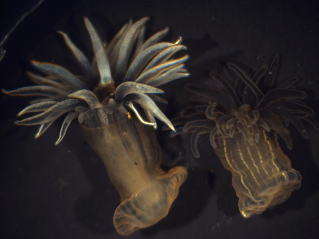
Under the stereomicroscope, Diadumene took on a new look.  The light colored tentacles had a luminescent glow, and with menthol anesthesia, the tentacles moved around in slow motion.   The anemones were partially immobilized, and the tentacles appeared shorter and more stout (compare the length of the tentacles of the anemones below to the ones in the photo above). Two color variants were seen, a majority with light yellow tentacles and a less frequent variant with light brown tentacles.

Diadumene lineata Growing on an Oyster Shell
                     
Diadumene lineata viewed with a stereomicroscope using fiber optic lighting.
The specimens were collected in July 2011 and anesthetized with menthol crystals.
The light colored tentacles had a luminescent glow.  2X zoom x 10X objective.

Diadumene lineata with Brown Translucent Tentacles
Diadumene lineata viewed from above showing the mouth and orange stripes.  This individual was also anesthetized with menthol crystals.  2X zoom x 10X objective.

Color Variants of Diadumene lineata
The two color variants of Diadumene lineata detached from the substrate
after menthol relaxation.  1.5X zoom x 10X objective.

View of the Mouth of Diadumene lineata
Beige colored mouth surrounded by rings of brown tentacles 
(above mouth, a small piece of adhering debris).  4X zoom x 10X objective.

Distribution of Diadumene in New England marinas (RI to Maine):
Pappal, A, Pederson, J, and Smith, JP.  Marine Invaders in the Northeast.  Rapid Assessment Survey of non-native and native marine species of floating dock communities. 7/25/2010 - 7/31/2010

Special thanks to Adrienne Pappal and Niels Hobbs for confirming the species identification of Diadumene from sterezoom micrographs.

Links providing further information on Diadumene lineata:

References on the history of Wellfleet:
1.  Wright, D.B. The Famous Beds of Wellfleet.  A Shellfishing History.The Wellfleet Historical Society, 153 pp., 2009. 
2.  Lombard, D.  Wellfleet, A Cape Cod Village.  Arcadia Publishing, 128 pp., 2000.  

Friday, December 9, 2011

New Ascidian Images on Tunicarium

Botryllus schlosseri is another beautiful colonial ascidian whose single and multi-color variants make for an amazing number of color morphs.  These colorful ascidans, like Botrylloides violaceus (Nov. 11 Post), make for terrific underwater photographs that I thought would transcend directly to the stereomicroscope.  It did to an extent; however, like with Botrylloides, structural detail was easier to capture with the lighter color variants.  Some of the stereomicrographs from this summer's monitoring of Cape Cod and Salem Sound marinas are now featured on Tunicarium, including images of the invasive ascidians Botrylloides violaceus, Botryllus schlosseri, and Didemnum vexillum.  

Botryllus schlosseri was a challenging colonial to capture under fiber optic lighting. The variants with dark-colored zooids blended in with the backgrounds making difficult the photography of structural detail.  With the light color variants against a darker background, the two-toned beige, which formed a mosaic-patterned colony, and single-colored orange colonies proved to be highly photogenic.

To capture the zooids with open siphons, I tried Gretchen Lambert's technique for relaxation with menthol crystals.  It was terrific for relaxing ascidians as well as other invertebrates.  But as with any approach, I found relaxation had its pros and cons for viewing as well as photomicrography.  Relaxation greatly reduced contraction ability, but also inhibited the ability of zooids to fully expand. Lack of anesthesia was challenging under bright light because individual zooids reacted to the light independently, each zooid in a cluster randomly closing and re-opening during photography.  My objective was to obtain images where all the branchial siphons in the field of view were fully open.  It made for some interesting and entertaining time-sequence shots.    

The images below are two of my favorites.  They demonstrate the difference between the information revealed capturing images of light vs. dark Botryllus color variants and show zooid structural and pigment cell details that cannot be seen solely by underwater photography.  The 40X original magnification generated images with the greatest resolution and zooid detail (4X zoom + 10X objective lens).  These details can also be seen out in the field with the 30-40X jeweler's loupe magnifying glasses that I discussed in the Nov. 25 Post.  The beige variant gave vivid zooid detail, whereas the typical white star on dark purple was outstanding for showing cells in the pigment bands.

Botryllus schlosseri Beige-Rust Mosaic Variant
Botryllus schlosseri growing on the green algae Codium fragile collected from MacMillan Wharf, Provincetown, MA. Zooids have a translucent beige color revealing the tips of the branchial tentacles and pale rust-orange cells in the pigment bands. 

Botryllus schlosseri White Star Variant with Parallel Pigment Bands.
Botryllus schlosseri growing on Codium collected from MacMillan Wharf, Provincetown, MA.  Four branchial openings are seen at the top with 4 pairs of white pigment bands leading to the single, common atrial aperture at the bottom center.  

The full collection of images on Tunicarium can be seen all together on Google Images that has been web-searched using the word "Tunicarium".  The photos are featured in thumbnail view and can be "double-clicked" to see the jpg files saved on the website.  Larger sized micrographs can be obtained from me by request at tomermak@rcn.com.

Friday, November 25, 2011

Helpful Monitoring Accessories

At the beginning of the summer, I got supplied with the usual set of monitoring supplies: Clipboard, Thermometer, Refractometer, Collection Trays, and a Pocket 6X Magnifying Glass.  I was all set, but when I arrived out in the field, the 6X magnification left me poorly equipped to make species determinations of the colonial species, and it was also difficult to distinguish the red bryozoan Bugula neritina from some filamentous red algae.  Back at home, under the stereomicroscope, I could easily make species determinations magnifying specimens 20-40X.  It was then that I realized I needed magnifiers at this range of magnification.
      
Off I went to the internet and found a terrific line of magnifiers at Amazon.  I purchased several, but the jeweler's loupes were the most helpful.  Some magnifiers are made for viewing details of photographs and the plane of focus is right against the glass, but the jeweler's loupes are designed to be held away from the subject and this feature keeps the lens clean and dry in the field.  

30X Jeweler's Loupe Magnifying Glass

The 20X, 30X, and 40X lenses are now permanent supplies in my monitoring bag; however I'd say that I use the 30X lens the most.  I use the 20X when I need a wider field of view and the 40X for the smallest details.  Nonetheless, I've used all of them on my most recent trips. 

Don't depend on these magnifying glasses to help much in image documentation, however.  Feeling in an adventurous spirit, I tried taking an iPhone photo of a small Botryllus schlosseri colony growing on Ulva.  I held the 30X jeweler's loupe lens directly against the iPhone lens, and surprisingly, I was able to obtain focus through the lens.  However, I could not hold the specimen or camera steady enough to take a sharp, in-focus photo.  My best attempt is shown below - not bad for evidence but insufficient for documentation.  At least the Botryllus was fresh!

A Blurry Botryllus schlosseri growing on Ulva Sea Lettuce
Photo taken with a 30X jeweler's loupe held directly against the camera lens
 of an iPhone mobile device. Time to get a macro camera........
  

Sunday, November 20, 2011

Convergence or Coincidence?

A Lighthearted Comparison & Identification Aid
                     
When identifying Botryllids, I frequently use the suburban housing development as a model for identifying Botrylloides.  This has been especially useful when the colonies are small or when the colors of Botrylloides violaceus and Botryllus schlosseri are similar. The organization of zooids in each species is distinctly different, with Botryllus forming stars and clusters and Botrylloides forming meandering chains of zooids reminiscent of newly constructed southwest housing tracts.

Human Social Organization - Shared Roads and Utilities.
   Botrylloides Organization - Shared Excurrent Canals and Blood Channels
 

It's fascinating when we see nature's designs show up in human social organization.  And somewhat validating, too, that we're all interconnected.

Friday, November 11, 2011

Marine Invasives Monitoring

My monitoring activities at Provincetown, Wellfleet, Salem, and Beverly Marinas are now completed for 2011 with the Species Reports for Ptown and Wellfleet sent off to the CZM and my marine biologic image collection significantly enlarged.  With 4 months of visits behind me, I obtained a nice selection of photographs of Massachusetts marinas and their marine invasive species (MIS).  After several of the trips, I was able to get brightfield microscopic images of colonial species within a few hours of their collection.  Fourteen of the sixteen species on the list in the August 8th Post were seen this season, all except the European oyster Ostrae edulis and leafy red alga Grateloupia turuturu.  Among the sessile or attached species, Didemnum vexillumStyela clavaBotrylloides violaceusBotryllus schlosseriDiplosoma listerianum, Ascidiella aspersaBugula neritinaMembranipora membranacea, and Codium fragile were commonly observed, whereas Diadumene lineata found a special niche in the Wellfleet Marina. 
             
The most dramatic event of the summer was the impact of Tropical Storm Irene which had a major erosive impact and removed most of the species that were not strongly attached or that extended out from the docks.  My visits to Cape Cod occurred 2-3 days after the storm, and the storm currents cleared away much of the overgrowth and removed some of the species down to the substrate. 
                                  
Perhaps the most impressive and beautiful species to my eye was Botrylloides violaceus, whose bright orange colors could be seen by casual observation walking on the docks and which looked even more beautiful up close when the meandering rows of zooids and their surrounding ampullae could be clearly observed.

Botrylloides violaceus in Clear View at Low Tide on a Pier Piling
 at Fisherman's Wharf, Provincetown, MA
The green alga Ulva lactuca grows above a band of orange Botrylloides violaceus. A colony of Didemnum vexillum can barely be seen below the Botrylloides.

Botrylloides violaceus Orange Colonies on the Sides of Dock Floats
View of the side of a dock at MacMillan Wharf, Provincetown, MA. Looking through the hazy water, light-orange, orange, brick-red-orange, and purple-red-orange color variants of Botrylloides can be distinguished. Also present were mature Mytilus edulis and Styela clava.

Botrylloides violaceus Colonial Organization 
Botrylloides colony showing meandering rows of zooids with open branchial siphons separated by clear tunic containing a border of amplullae.  3X zoom x 10X objective.

Botrylloides violaceus Zooids and Ampullae 
Orange color variant of Botrylloides showing branchial siphons, pigmented body wall, and pigmented ampullae between rows of zooids. Endostyle is in subtle contrast to body wall.
3X zoom x 10X objective.

Botrylloides violaceus Endostyle and Branchial Features
Light-orange color variant showing branchial tentacles and orange-pigmented endostyle against the more transparent body wall.  3X zoom x 10X objective.
        
Additional stereozoom micrographs of Botrylloides violaceus and other ascidians can be seen at http://www.tunicarium.com/.